Jocelyn Nestle
  • Blog
  • About
  • Contact

Fallen Arches Causes, Conditions And Treatment Method

7/4/2017

 
Overview

Adult Acquired Flat Foot

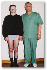
Fallen arches is one way of describing a flat or pronated foot. This can sometimes be implicated in ongoing problems such as lower back pain, knee pain, ankle pain, shin pain etc. The reason for the pronated foot is often due to the alignment of the bones within the foot, but can be increased by such things as ligament laxity, high body weight and a number of other specific conditions. One of these is posterior tibial dysfunction. This is a progressive disorder which allows the foot to pronate or lower on the inside section and can, if left untreated, result in quite debilitating effects.

Causes

There are several factors that can contribute to the development of fallen arches. These factors include the following. Genetic abnormality, torn or stretched tendons, amage to the posterior tibial tendon, bone fractures, dislocation of bones, nerve damage, rheumatoid arthritis and other medical conditions. In addition, there are other factors that can increase your risk of developing fallen arches. These risk factors include diabetes, pregnancy, Obesity and Aging.

Symptoms

The primary symptom of flatfeet is the absence of an arch upon standing. Additional signs of flatfeet include the following. Foot pain. Pain or weakness in the lower legs. Pain or swelling on the inside of the ankle. Uneven shoe wear. While most cases of flatfeet do not cause problems, complications can sometimes occur. Complications include the following, bunions and calluses, inability to walk or run normally, inflammation and pain in the bottom of the foot (plantar fasciitis), tendonitis in the Achilles heel and other ligaments, pain in the ankles, knees, and hips due to improper alignment, shin splints, stress fractures in the lower legs.

Diagnosis

Podiatrists are trained in expertly assessing flat feet and identifying different risk factors and the causes for it. Initial assessment will begin with a detailed history attempting to find out if any underlying illness has resulted in this. A detailed clinical examination normally follows. The patient may be asked to perform certain movements such as walking or standing on their toes to assess the function of the foot. Footwear will also be analysed to see if there has been excessive wear or if they are contributing to the pronation of the foot. To assess the structure of the foot further, the podiatrist may perform certain x-rays to get a detailed idea of the way the bones are arranged and how the muscle tissues may be affecting them. It also helps assess any potential birth defects in a bit more detail.

fallen arches support

Non Surgical Treatment

Treatment of flat feet by a fully educated, trained, and licensed podiatrist should be sought if the previously mentioned pain in the foot or knee areas start to surface and becomes a painful problem. Depending on the cause and exact type of the condition, a variety of forms of treatment may be prescribed. Verification of the exact cause and type of flat feet that each individual has, should only be handled by a professional podiatrist. Often times, the actual treatment method will include some form of arch support or light gymnastic style exercise. In rare cases, something more involved may be necessary, but only your skilled podiatrist would be able to accurately make that determination. If you suffer from flat feet, plantar fasciitis, or any other form of problematic foot condition, we encourage you to contact a foot clinic today and potentially rid yourself of that problem in virtually no time at all.

Surgical Treatment

Adult Acquired Flat Feet

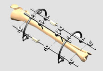
This is rare and usually only offered if patients have significant abnormalities in their bones or muscles. Treatments include joint fusion, reshaping the bones in the foot, and occasionally moving around tendons in the foot to help balance out the stresses (called tendon transfer).

Leg Length Discrepancy Heel Lifts

6/30/2017

 
Overview

Surgery to shorten the longer leg. This is less involved than lengthening the shorter leg. Shortening may be done in one of two ways. Closing the growth plate of the long leg 2-3 years before growth ends (around age 11-13), letting the short leg catch up. This procedure is called an epiphysiodesis. Taking some bone from the longer leg once growth is complete to even out leg lengths. Surgery to lengthen the shorter leg. This surgery is more involved than surgery to shorten a leg. During this surgery, cuts are made in the leg bone. An external metal frame and bar are attached to the leg bone. This frame and bar slowly pull on the leg bone, lengthening it. The frame and bar must be worn constantly for months to years. When the frame and bar are removed, a leg cast is required for several months. This surgery requires careful and continued follow-up with the surgeon to be sure that healing is going well.Leg Length Discrepancy

Causes

Some children are born with absence or underdeveloped bones in the lower limbs e.g., congenital hemimelia. Others have a condition called hemihypertrophy that causes one side of the body to grow faster than the other. Sometimes, increased blood flow to one limb (as in a hemangioma or blood vessel tumor) stimulates growth to the limb. In other cases, injury or infection involving the epiphyseal plate (growth plate) of the femur or tibia inhibits or stops altogether the growth of the bone. Fractures healing in an overlapped position, even if the epiphyseal plate is not involved, can also cause limb length discrepancy. Neuromuscular problems like polio can also cause profound discrepancies, but thankfully, uncommon. Lastly, Wilms? tumor of the kidney in a child can cause hypertrophy of the lower limb on the same side. It is therefore important in a young child with hemihypertrophy to have an abdominal ultrasound exam done to rule out Wilms? tumor. It is important to distinguish true leg length discrepancy from apparent leg length discrepancy. Apparent discrepancy is due to an instability of the hip, that allows the proximal femur to migrate proximally, or due to an adduction or abduction contracture of the hip that causes pelvic obliquity, so that one hip is higher than the other. When the patient stands, it gives the impression of leg length discrepancy, when the problem is actually in the hip.

Symptoms

The effects of a short leg depend upon the individual and the extent of discrepancy. The most common manifestation if a lateral deviation of the lumbar spine toward the short side with compensatory curves up the spine that can extend into the neck and even impacts the TMJ. Studies have shown that anterior and posterior curve abnormalities also can result.

Diagnosis

The doctor carefully examines the child. He or she checks to be sure the legs are actually different lengths. This is because problems with the hip (such as a loose joint) or back (scoliosis) can make the child appear to have one shorter leg, even though the legs are the same length. An X-ray of the child?s legs is taken. During the X-ray, a long ruler is put in the image so an accurate measurement of each leg bone can be taken. If an underlying cause of the discrepancy is suspected, tests are done to rule it out.

Non Surgical Treatment

The object of treatment for leg length discrepancy is to level the pelvis and equalize the length of the two limbs. Inequalities of 2-2.5 centimeters can be handled with the following. Heel lifts/ adjustable heel lifts can be used inside a shoe where shoes have a full heel counter. Heel lifts may be added to the heel on the outside of the shoe along with an internal heel lift. Full platforms along the forefoot and rearfoot area of a shoe can be added. There are many different adjustable heel lifts available on the market. For treatment of a leg length discrepancy, consult your physician. They may refer you to a Physiotherapist or Chiropractor for determination of the type of LLD. A Certified Pedorthist (Canada) will treat a structural leg length discrepancy with a heel lift or in larger discrepancies a footwear modification.

Leg Length

height increase exercises

Surgical Treatment

Your child will be given general anesthetic. We cut the bone and insert metal pins above and below the cut. A metal frame is attached to the pins to support the leg. Over weeks and months, the metal device is adjusted to gradually pull the bone apart to create space between the ends of the bones. New bone forms to fill in the space, extending the length of the bone. Once the lengthening process is completed and the bones have healed, your child will require one more short operation to remove the lengthening device. We will see your child regularly to monitor the leg and adjust the metal lengthening device. We may also refer your child to a physical therapist to ensure that he or she stays mobile and has full range of motion in the leg. Typically, it takes a month of healing for every centimeter that the leg is lengthened.

Heel Soreness The Main Causes, Warning Signs And Cure Possibilities

6/28/2017

 
Overview

Pain At The Heel

The most common cause of heel pain is plantar fasciitis. Many patients with plantar fasciitis have a heel spur on the front and bottom of their heel, but heel spurs do not cause pain. The common name is "heel spur" because it's easier to pronounce than "plantar fasciitis" and doctors are able to point to the spur on an x-ray. Causes of heel pain include inadequate flexibility in the calf muscles, lack of arch support, being overweight, suddenly increasing activity, and spending too much time on the feet. Arch support was rated the best treatment in our first survey of 1,800 visitors to heelspurs.com. The Plantar Fasciitis Orthotic is getting the best customer reviews and it is the most popular product. It is new for 2011. Returning customers may be seeking the Pinnacle Orthotic. The primary difference is that the PF Orthotic should be used only for current cases of plantar fasciitis or heel spurs and the Pinnacle is best for general use once the condition has subsided. Survery respondents also benefited from: rest, ice, tape, and night splints.

Causes

In the majority of cases, heel pain has a mechanical cause. It may also be caused by arthritis, infection, an autoimmune problem trauma, a neurological problem, or some other systemic condition (condition that affects the whole body).

Symptoms

See your doctor as soon as possible if you experience severe pain accompanied by swelling near your heel. There is numbness or tingling in the heel, as well as pain and fever. There is pain in your heel as well as fever. You are unable to walk normally. You cannot bend your foot downwards. You cannot stand with the backs of the feet raised (you cannot rise onto your toes). You should arrange to see a doctor if the heel pain has persisted for more than one week. There is still heel pain when you are not standing or walking.

Diagnosis

The diagnosis of heel pain and heel spurs is made by a through history of the course of the condition and by physical exam. Weight bearing x-rays are useful in determining if a heel spur is present and to rule out rare causes of heel pain such as a stress fracture of the heel bone, the presence of bone tumors or evidence of soft tissue damage caused by certain connective tissue disorders.

Non Surgical Treatment

If pain and other symptoms of inflammation-redness, swelling, heat-persist, you should limit normal daily activities and contact our office, or another doctor of podiatric medicine. Your foot would be examined, and an X-ray may be taken to rule out problems of the bone. Early treatment might involve oral or injectable anti-inflammatory medication, taping, padding, massage, stretching, exercise, shoe recommendations, physiotherapy, over-the-counter shoe inserts or, if the condition is chronic and there is a biomechanical basis to the complaint, orthoses (or orthotic devices) may be used to permanently take strain off the fascia. Only rarely is surgery required for heel pain. If necessary, however, it may involve the release of the plantar fascia, removal of a spur, removal of a bursa, or removal of a neuroma or other soft-tissue growth.

Surgical Treatment

Surgery to correct heel pain is generally only recommended if orthotic treatment has failed. There are some exceptions to this course of treatment and it is up to you and your doctor to determine the most appropriate course of treatment. Following surgical treatment to correct heel pain the patient will generally have to continue the use of orthotics. The surgery does not correct the cause of the heel pain. The surgery will eliminate the pain but the process that caused the pain will continue without the use of orthotics. If orthotics have been prescribed prior to surgery they generally do not have to be remade.

bestshoelifts

Prevention

Heel Pain

Being overweight can place excess pressure and strain on your feet, particularly on your heels. Losing weight, and maintaining a healthy weight by combining regular exercise with a healthy, balanced diet, can be beneficial for your feet. Wearing appropriate footwear is also important. Ideally, you should wear shoes with a low to moderate heel that supports and cushions your arches and heels. Avoid wearing shoes with no heels.

Leg Length Discrepancy Following Knee Replacement Surgery

6/26/2017

 
Overview

The bone is lengthened by surgically applying an external fixation device to the leg. The external fixator, a scaffold-like frame, is connected to the bone with wires, pins, or both. A small crack is made in the bone and the frame creates tension when the patient or family member turns its dial. This is done several times each day. The lengthening process begins approximately five to 10 days after surgery. The bone may lengthen 1 millimeter per day, or approximately 1 inch per month. Lengthening may be slower in a bone that was previously injured. It may also be slower if the leg was operated on before. Bones in patients with potential blood vessel abnormalities, such as cigarette smokers, may also need to be lengthened more slowly. The external fixator is worn until the bone is strong enough to support the patient safely. This usually takes about three months for each inch. Factors such as age, health, smoking and participation in rehabilitation can affect the amount of time needed.Leg Length Discrepancy

Causes

Leg length discrepancies can be caused by: hip and knee replacements, lower limb injuries, bone diseases, neuromuscular issues and congenital problems. Although discrepancies of 2 cm or less are most common, discrepancies can be greater than 6 cm. People who have LLD tend to make up for the difference by over bending their longer leg or standing on the toes of their shorter leg. This compensation leads to an inefficient, up and down gait, which is quite tiring and over time can result in posture problems as well as pain in the back, hips, knees and ankles.

Symptoms

As patients develop LLD, they will naturally and even unknowingly attempt to compensate for the difference between their two legs by either bending the longer leg excessively or standing on the toes of the short leg. When walking, they are forced to step down on one side and thrust upwards on the other side, which leads to a gait pattern with an abnormal up and down motion. For many patients, especially adolescents, the appearance of their gait may be more personally troublesome than any symptoms that arise or any true functional deficiency. Over time, standing on one's toes can create a contracture at the ankle, in which the calf muscle becomes abnormally contracted, a condition that can help an LLD patient with walking, but may later require surgical repair. If substantial enough, LLD left untreated can contribute to other serious orthopaedic problems, such as degenerative arthritis, scoliosis, or lower back pain. However, with proper treatment, children with leg length discrepancy generally do quite well, without lingering functional or cosmetic deficiencies.

Diagnosis

A qualified musculoskeletal expert will first take a medical history and conduct a physical exam. Other tests may include X-rays, MRI, or CT scan to diagnose the root cause.

Non Surgical Treatment

In an adult, we find that we can add a non compressive silicone heel lift to a shoe in increments of 3-4 mm maximum per week. Were we to give a patient with a 20 mm short leg, 20 mm of lift all at once, their entire body would rebel. The various compensations that the body has made, such as curvatures and shortening of muscles on the convex side of the curve, would make such a dramatic change not just noticeable, but painful. When we get close to balancing a patient by lifting a leg with heel inserts, then we perform another gait analysis and follow up xray. At that point, we can typically write them a final prescription to have their shoe modified. A heel lift is typically fine up to 7 mm. When it gets higher than that, the entire shoe must be modified. There are two reasons for this. The back of the shoe is generally too short to accommodate more than 7-8 mm inserted inside the shoes and a heel lift greater than 7 mm will lead to Achilles tendon shortening, which then creates it?s own panoply of problems.

Leg Length

how to increase height naturally after 18

Surgical Treatment

Surgical operations to equalize leg lengths include the following. Shortening the longer leg. This is usually done if growth is already complete, and the patient is tall enough that losing an inch is not a problem. Slowing or stopping the growth of the longer leg. Growth of the lower limbs take place mainly in the epiphyseal plates (growth plates) of the lower femur and upper tibia and fibula. Stapling the growth plates in a child for a few years theoretically will stop growth for the period, and when the staples were removed, growth was supposed to resume. This procedure was quite popular till it was found that the amount of growth retarded was not certain, and when the staples where removed, the bone failed to resume its growth. Hence epiphyseal stapling has now been abandoned for the more reliable Epiphyseodesis. By use of modern fluoroscopic equipment, the surgeon can visualize the growth plate, and by making small incisions and using multiple drillings, the growth plate of the lower femur and/or upper tibia and fibula can be ablated. Since growth is stopped permanently by this procedure, the timing of the operation is crucial. This is probably the most commonly done procedure for correcting leg length discrepancy. But there is one limitation. The maximum amount of discrepancy that can be corrected by Epiphyseodesis is 5 cm. Lengthening the short leg. Various procedures have been done over the years to effect this result. External fixation devices are usually needed to hold the bone that is being lengthened. In the past, the bone to be lengthened was cut, and using the external fixation device, the leg was stretched out gradually over weeks. A gap in the bone was thus created, and a second operation was needed to place a bone block in the gap for stability and induce healing as a graft. More recently, a new technique called callotasis is being use. The bone to be lengthened is not cut completely, only partially and called a corticotomy. The bone is then distracted over an external device (usually an Ilizarov or Orthofix apparatus) very slowly so that bone healing is proceeding as the lengthening is being done. This avoids the need for a second procedure to insert bone graft. The procedure involved in leg lengthening is complicated, and fraught with risks. Theoretically, there is no limit to how much lengthening one can obtain, although the more ambitious one is, the higher the complication rate.

Mortons Neuroma Treatment

6/6/2017

 
Overview

Morton
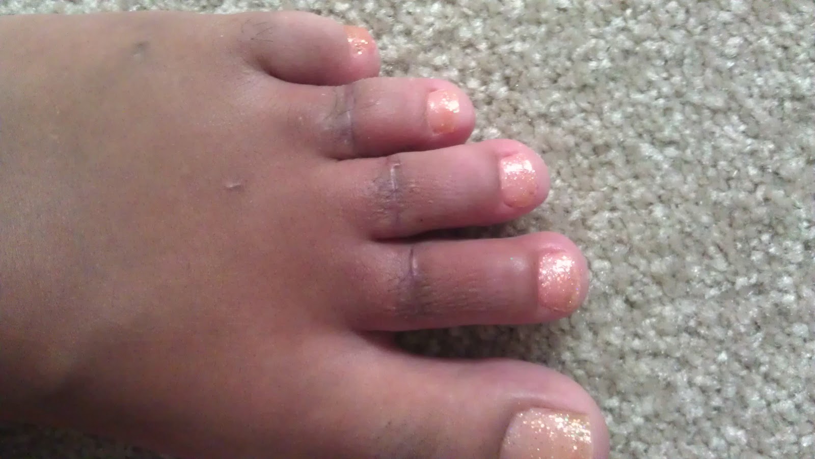
Morton neuroma (interdigital neuroma), first described in 1876, is a perineural fibrosis and nerve degeneration of the common digital nerve. Morton neuroma, or Morton's neuroma, is not a true neuroma, although it results in neuropathic pain in the distribution of the interdigital nerve secondary to repetitive irritation of the nerve. The most frequent location is between the third and fourth metatarsals (third webspace). Other, less common locations are between the second and third metatarsals (second webspace) and, rarely, between the first and second (first webspace) or fourth and fifth (fourth webspace) metatarsals.

Causes

Wearing shoes that are too tight can make the pain of Morton's neuroma worse. This is because the toe bones are more likely to press on the affected nerve if your shoes are too tight. High-heeled shoes, particularly those over 5cm (2 inches), or shoes with a pointed or tight toe area, can also compress your toes and make the pain worse. This is why women tend to be affected by Morton's neuroma more than men.

Foot conditions.

Symptoms

If you have a Morton?s neuroma, you may have one or more of these symptoms where the nerve damage is occurring, Tingling, burning, or numbness, pain, a feeling that something is inside the ball of the foot or a feeling that there?s something in the shoe or a sock is bunched up. The progression of a Morton?s neuroma often follows the following pattern. The symptoms begin gradually. At first they occur only occasionally, when wearing narrow-toed shoes or performing certain aggravating activities. The symptoms may go away temporarily by removing the shoe, massaging the foot, or by avoiding aggravating shoes or activities. Over time the symptoms progressively worsen and may persist for several days or weeks. The symptoms become more intense as the neuroma enlarges and the temporary changes in the nerve become permanent.

Diagnosis

Morton's neuroma is usually diagnosed by your doctor listening to your symptoms and examining your foot. Sometimes your doctor can feel the 'neuroma', or an area of thickening in your foot, which may be tender. Sometimes, your doctor may suggest an ultrasound scan or MRI scan to confirm the diagnosis but this is not always necessary. Some doctors inject a local anaesthetic into the area where you are experiencing pain. If this causes temporary relief of pain, burning and tingling, it can sometimes help to confirm the diagnosis and show the doctor where the problem is.

Non Surgical Treatment

Initial treatment for Morton?s Neuroma may include non-prescription anti-inflammatory medications to reduce pain and swelling. These may consist of standard analgesics such as aspirin and ibuprofen (Advil, Motrin, others). Massaging the painful region three times daily with ice. Change of footwear. Avoid tight shoes, high heels or any footwear that seems to irritate the condition. Low heeled shoes with softer soles are preferable. Arch supports and foot pads to help reduce pressure on the nerve. In some cases, a physician may prescribe a customized shoe insert, molded to fit the contours of the patient?s foot. Reducing activities causing stress to the foot, including jogging, dancing, aerobic activity or any high impact movements of the foot. Injections of a corticosteroid medication to reduce the swelling and inflammation of the nerve and reduce pain. Occasionally other substances may be injected in order to ?ablate? the Neuroma. (The overuse of injected steroids is to be avoided however, as side effects, including weight gain and high blood pressure can result.)Morton

Surgical Treatment

Should the problem have progressed beyond the point where these measures are sufficient, the podiatric professional may recommend surgery. This procedure involves excision of the involved nerve mass. This will relieve the pain. Many patients report permanent numbness in the spot formerly affected by the neuroma, but prefer it to the pain. Most surgeries are successful; unfortunately, there are cases where the patient suffers another neuroma, sometimes in or near the same spot as before. A podiatric professional can explain the statistics of recurrence in various cases. If you suspect a neuroma, don?t wait for it to get better on its own. The earlier the diagnosis, the higher the likelihood that it can be treated with conservative measures. Don?t think that foot pain of any kind is inevitable, either, even if it runs in your family.

Alleviating Leg Length Discrepancy With Shoe Lifts

2/20/2016

 
There are actually not one but two different types of leg length discrepancies, congenital and acquired. Congenital implies you are born with it. One leg is structurally shorter compared to the other. As a result of developmental phases of aging, the brain senses the gait pattern and identifies some variance. Your body typically adapts by dipping one shoulder to the "short" side. A difference of under a quarter inch isn't really irregular, doesn't need Shoe Lifts to compensate and ordinarily doesn't have a serious effect over a lifetime.

Leg Length Discrepancy Shoe Lifts

Leg length inequality goes typically undiagnosed on a daily basis, however this problem is simply corrected, and can eradicate a number of cases of low back pain.

Therapy for leg length inequality usually consists of Shoe Lifts. These are generally very reasonably priced, usually priced at under twenty dollars, compared to a custom orthotic of $200 or higher. When the amount of leg length inequality begins to exceed half an inch, a whole sole lift is generally the better choice than a heel lift. This prevents the foot from being unnecessarily stressed in an abnormal position.

Lower back pain is easily the most common ailment affecting people today. Around 80 million men and women suffer from back pain at some point in their life. It is a problem which costs companies millions annually because of time lost and output. Fresh and superior treatment methods are constantly sought after in the hope of minimizing the economical influence this issue causes.

Shoe Lifts

People from all corners of the earth suffer from foot ache as a result of leg length discrepancy. In these types of situations Shoe Lifts can be of beneficial. The lifts are capable of alleviating any pain and discomfort in the feet. Shoe Lifts are recommended by many specialist orthopaedic doctors.

In order to support the human body in a well balanced manner, the feet have a critical role to play. Despite that, it is often the most overlooked zone in the body. Some people have flat-feet meaning there may be unequal force exerted on the feet. This causes other areas of the body like knees, ankles and backs to be impacted too. Shoe Lifts ensure that proper posture and balance are restored.

Flexible Hammertoes Treatment

7/21/2015

 
HammertoeOverview

A Hammer toe is a deformity of the middle joint of a toe, producing a clenched, clawlike appearance in the affected digit. The tendons in the toe become abnormally contracted, causing the toe to bend downward, which, in turn, forces the joint to protrude upward. A mallet toe is a deformity in which the end joint of a toe becomes bent downward, so that the toe curls underneath itself. In either case the affected joints are stiff, and often the toe cannot be straightened out. Constant rubbing against shoes may furthermore cause a painful corn (a round patch of rough, thickened, calloused skin) to develop over the joint or at the tip of the affected toe. Hammer and mallet toes may occur in any toe, although the second toe is the most common site. These deformities are often painful and limit the toe?s range of motion-sometimes requiring surgery.

Causes

Hammer toe usually affects the second toe. However, it may also affect the other toes. The toe moves into a claw-like position. The most common cause of hammer toe is wearing short, narrow shoes that are too tight. The toe is forced into a bent position. Muscles and tendons in the toe tighten and become shorter. Hammer toe is more likely to occur in women who wear shoes that do not fit well or have high heels and children who keep wearing shoes they have outgrown. The condition may be present at birth (congenital) or develop over hammertoes time. In rare cases, all of the toes are affected. This may be caused by a problem with the nerves or spinal cord.

Hammer ToeSymptoms

Pain upon pressure at the top of the bent toe from footwear. The formation of corns on the top of the joint. Redness and swelling at the joint contracture. Restricted or painful motion of the toe joint. Pain in the ball of the foot at the base of the affected toe.

Diagnosis

The treatment options vary with the type and severity of each hammer toe, although identifying the deformity early in its development is important to avoid surgery. Your podiatric physician will examine and X-ray the affected area and recommend a treatment plan specific to your condition.

Non Surgical Treatment

Hammer toes may be effectively corrected in different ways. Treatments can be non-invasive and involve physical therapy along with the advice that the person not wear any more shoes that restrict appropriate space for their toes. Appropriate shoes for people who want to avoid hammer toes, or for people who already have them, should be at least half an inch longer than the person's longest toe. High-heeled shoes are something to definitely avoid.

Surgical Treatment

Toe Relocation procedures are ancillary procedures that are performed in conjunction with one of the two methods listed about (joint resection or joint mending). When the toe is deformed (buckled) at the ball of the foot, then this joint often needs to be re-positioned along with ligament releases/repair to get the toe straight. A temporary surgical rod is needed to hold the toe aligned while the ligaments mend.

Hammer ToePrevention

The best treatment is good prevention! Hammertoe can be prevented by wearing shoes with ample toe room, avoiding high heels, and wearing adjustable shoes to assure a looser fit. When buying shoes, shop at the end of the day when your feet are swollen from daily activity, try both shoes on to confirm they fit properly, and if necessary, visit a shoe repair store to see if they can stretch your shoes for a better fit.

How To Treat Tailor'S Bunions

6/26/2015

 
Overview
Bunions Callous If you have bunions, taking care of them now can help you avoid more serious treatment later. A bunion is a bump that forms on the joint of the big toe when bone or tissue moves out of place and extends beyond the normal anatomy of the toe. Left untreated, bunions can cause debilitating pain and may require surgery to correct.

Causes
Women tend to suffer from bunions more often than men, probably because of the shoes they wear. But besides shoes, standing on the feet for long periods of time can also make symptoms of a bunion worse. Along with the bump, these bunion symptoms include pain or soreness, swelling, redness around the joint, a burning sensation or sometimes numbness. The big toe may develop calluses or not be able to move as well as it once did. Sores between the toes and ingrown toenails may also occur because of a bunion.

Symptoms
Often the bunion is not painful and the individual leads a normal active life. Other times the bunion can be very painful, even debilitating. Pain is usually very achy and typically radiates to the toes and along the arch of the foot. Due to the abnormal positioning of the bones in the foot, sharp nerve pains could also be present.

Diagnosis
X-rays are the best way to determine the amount of deformity of the MTP joint. Blood work may be required to rule out other diseases that may be associated with bunions such as rheumatoid arthritis. Other tests such as bone scans or MRI's are not usually required.

Non Surgical Treatment
Most of the time, non-surgical (conservative) treatment can control the symptoms of a foot bunion or bunionette. These include. Appropriate Footwear, changing to wide fitting footwear reduces the pressure on the big toe and prevents shoes from rubbing on the bony lump. When possible, go barefoot Toe stretchers are a really simple way to reduce foot bunion pain. Toe Stretchers, wearing toe spaces that fit in-between the toes help to stretch the muscles and ligaments around the toes, improving the alignment and relieving pain. Find out more about how these work in the Toe Stretcher section. Painkillers, your doctor may prescribe or recommend over-the-counter medication to reduce the pain and inflammation. Foot bunion correctors can be worn in your shoe to help realign your foot if you suffer from foot bunions. Orthotics. There are a number of over-the-counter shoe inserts that can help relieve symptoms. Bunion correctors work by realigning the bones in your foot to reduce pressure on the affected toe. There are both day-time and night-time splints on the market, although the evidence of their effectiveness is lacking. Ice. Applying ice packs to the foot can help reduce pain and inflammation. Bunion pads help to reduce any friction on your big toe. Bunion Pads. You can also get protective foot cushions that sit over the skin to prevent the hallux abducto valgus rubbing on your shoes. Bunion Pain

Surgical Treatment
If non-surgical treatments have failed to relieve your bunion pain, or when the pain is interfering with your daily activities, contact the Dallas bunion surgery specialists at North Texas Foot & Ankle to discuss surgical options. There are a several ways to perform bunion surgery. The best procedure for one person is not necessarily the best for another. Some procedures allow you to walk much sooner, reducing the need for crutches. Depending on your foot type, the procedure can have a greater risk for return of the bunion deformity. Other procedures may require you to be on crutches for a few weeks - but could offer a better result in the long-term. Learn more about the different types of bunion surgery.

Prevention
To help prevent bunions be sure your shoes don't cramp or irritate your toes. Choose shoes with a wide toe box - there should be space between the tip of your longest toe and the end of the shoe. Your shoes should conform to the shape of your feet without squeezing or pressing any part of your foot. Avoid pointy-toed shoes.
Powered by Create your own unique website with customizable templates.